For some people, using an oxygen breathing machine at home is the best way to effectively treat the effects of lung disease or COPD, allowing them to breathe better. If oxygen therapy is required to treat your COPD, your doctor will prescribe a respiratory oxygen machine. Your health care providers will decide how frequently breathing machines for COPD should be used for your oxygen therapy, how much oxygen should be consumed (liters per minute) and when you should use your breathing machine. A simple blood test will confirm the right prescription for your oxygen therapy for COPD. Some people with COPD will need oxygen therapy only when doing something strenuous, such as during exercise or extended periods of walking. Other COPD patients may only need oxygen therapy for at night. Many patients prescribed oxygen therapy however, need oxygen continuously—day, night and when traveling—and will need to find a versatile solution to provide portable oxygen for COPD and fit their lifestyle. [1,2] According to the American Lung Association, supplemental oxygen improves sleep, mood, mental alertness, and stamina, and allows individuals to carry out normal, everyday functions. [2]
Below are the different solutions available for oxygen therapy delivery, as well as the advantages and challenges of oxygen for breathing problems with each system. Your doctor will provide guidelines for you to help you enjoy the full benefits of oxygen therapy in COPD. Discover how being a COPD patient on oxygen therapy can work with your life.
Oxygen Therapy for COPD: Three Types of Oxygen Delivery Systems

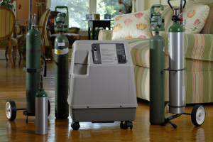
One drawback to compressed gas tanks is that you must always have enough tanks on hand so you do not run out of oxygen, which means managing frequent visits from your oxygen provider and finding a place to store larger tanks for home use and portable oxygen tanks for breathing while you are out.[4] If you are a COPD patient and oxygen therapy has been prescribed for you, compressed gas is likely what you have pictured using. However, it may not be your ideal solution for portable oxygen.
Liquid Oxygen
Liquid oxygen is a type of oxygen delivery where oxygen is compressed and cooled to a point that it becomes frozen. Most liquid oxygen systems provide a high concentration oxygen and do not require any electricity to run. They usually consist of a stationary storage unit and a portable container. Liquid oxygen is very cold (-297° F) and can cause frostbite burns to the skin if not handled with care, especially when filling a portable tank. A liquid oxygen system consists of a stationary unit and a portable device. Liquid oxygen is very cold and can cause frostbite or burns if it comes in direct contact with your skin. When the liquid is released from the container, it changes back to a gas as it warms up so that you are able to breathe it.[4] Liquid oxygen evaporates over time so don’t fill tanks too far ahead of when you need to use them.[5]
There are some drawbacks with using liquid oxygen for your oxygen therapy for COPD. While the liquid oxygen delivery system is smaller than the traditional compressed canister system, liquid oxygen is also more expensive, which can be a hardship for patients requiring long-term oxygen therapy for lung disease. Additionally, you must be very careful using this equipment, and particularly when transferring the liquid oxygen from the large tank to the small oxygen tank for breathing to avoid injury from the dangerously cold liquid oxygen. Because of the cost and the care that must be taken when using liquid oxygen for oxygen therapy for COPD, it is not always the ideal choice for both the COPD patient and oxygen therapy.[3]
Oxygen Concentrators

Single Solution Oxygen Concentrators
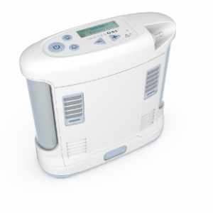
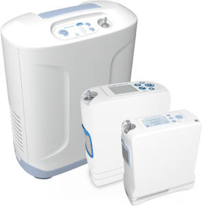
This is a newer technology, in which a portable oxygen concentrator is capable of satisfying all of your oxygen therapy for COPD needs, including stationary, portable and travel use. The Inogen One System is capable of continuous use in a home, institution, vehicles and various mobile environments. It is used on a prescriptive basis by patients requiring supplemental oxygen including but not restricted to those with chronic obstructive pulmonary disease (COPD).
Read on to learn more about Inogen One portable oxygen concentrators and how they can help with your oxygen therapy for COPD.
The Inogen One System Is Designed for Oxygen Therapy for COPD
The benefits of oxygen therapy in COPD are greatly increased when you can allow increased daily oxygen use and/or better compliance with portable oxygen for COPD.[2] Each portable oxygen concentrator model in the Inogen One System is designed to be used at home or on the go, all day, every day.
Inogen’s purpose is to improve lives through respiratory therapy, so if you are a COPD patient and oxygen therapy is in your future, talk to your doctor about whether a portable oxygen concentrator from Inogen is right for your needs.
Oxygen Therapy for COPD Guidelines: Safety and Oxygen Therapy [2]
Although oxygen is a safe, non-flammable gas, it does support combustion—in other words, some materials can readily catch fire and burn in the presence of oxygen. For that reason, just as with any medical treatment, it’s important to follow certain precautions while using it.
If you or a loved one is prescribed supplemental oxygen therapy, stay safe by:
- Storing oxygen properly: Oxygen canisters should be kept upright and in a place where they won’t be able to fall over or roll; an oxygen storage cart or similar device is ideal. Store canisters well away from any type of heat source, gas stove, or lit candles.
- Posting “no smoking” signs around your home to remind visitors not to smoke near you or your oxygen.
- Using caution around open flames like matches and candles, as well as gas heaters and stoves. If you are using supplemental oxygen, you should be at least five feet away from all heat sources.
- Turning off oxygen supply valves when not in use.
Post the phone number of the company that makes your oxygen canisters and other supplies in a visible location in case you have any questions about the equipment.
And in the event of a fire, make sure you know how to properly use a fire extinguisher. Accidents can happen, but don’t need to be tragic if you’re prepared.
Potential Side Effects of Oxygen Therapy
It is helpful to be aware of potential side effects COPD patients could experience during your oxygen treatments. Make sure to always follow your healthcare provider’s direction. Here is what to watch for:
Nasal dryness and skin irritation: The most common side effect of using long-term supplemental oxygen is nasal dryness and skin irritation, primarily in the places where the cannula and tubing touches the face. Use a humidifier at home or saline solution to make nasal passages less dry, and be sure to take care of your skin by applying lotions as needed to prevent irritation. [6]
Oxygen toxicity – When it comes to oxygen therapy, there can be too much of a good thing. Prolonged administration of highly concentrated oxygen can potentially damage the lung lining tissues and air sacs, a condition known as pulmonary oxygen toxicity. The risk for this side effect of oxygen therapy comes into play primarily when patients on home treatment increase the oxygen flow rate to a level higher than prescribed. [6] Always follow the prescription provided by your healthcare provider.
Breathing suppression: The brain respiratory center monitors levels of oxygen and carbon dioxide in the blood. The level of carbon dioxide in the blood normally serves as the primary stimulant for the breathing. In some patients with chronic obstructive pulmonary disease, or COPD, the blood oxygen level becomes the primary stimulator of the respiratory rate, a phenomenon known as hypoxic respiratory drive. Starting patients with hypoxic respiratory drive on a low concentration of supplemental oxygen typically helps avoid this potentially serious side effect of oxygen therapy. [6]
Talk to your doctor about any side effects from oxygen therapy you experience and do not make any adjustments to your oxygen therapy without your doctor’s permission.
Contact Inogen today to find out more about how our portable oxygen concentrators can provide oxygen therapy for COPD.
Frequently Asked Questions: Oxygen Therapy for COPD
Why do patients with COPD need oxygen?
COPD is a chronic, progressive lung disease that causes damage to the lungs. The result of this damage is that the lungs are unable to absorb oxygen properly, which leaves patients oxygen deprived. Oxygen therapy for COPD increases the amount of oxygen available to patients, allowing them to absorb more oxygen and improve their oxygen saturation.
Oxygen therapy for COPD side effects helps treat the symptoms of COPD, reducing shortness of breath, discomfort and fatigue. The benefits of oxygen therapy in COPD are quite extensive, which is why it is a common treatment for this lung disease. [1,2]
How much oxygen should be given to a patient with COPD?
Only your doctor can decide what your oxygen prescription should be, which will include the amount of time and frequency that you receive oxygen therapy for COPD side effects, as well as the flow rate of your oxygen for breathing problems. Generally speaking, the goal is to get your oxygen saturation levels up to above 90%, but as a COPD patient and oxygen therapy user, you may also be at an elevated risk of hyperoxia (too much oxygen) as well as the potential risk of retaining too much carbon dioxide.[7] As such, it is essential that you follow your doctor’s prescription and never adjust your oxygen therapy for COPD without speaking to your health care provider first.
What happens when COPD patients get too much oxygen?
When it comes to oxygen therapy, there can be too much of a good thing. Prolonged administration of highly concentrated oxygen can potentially damage the lung lining tissues and air sacs, a condition known as pulmonary oxygen toxicity. The risk for this side effect of oxygen therapy comes into play primarily when patients on home treatment increase the oxygen flow rate to a level higher than prescribed. [5] Always follow the prescription provided by your healthcare provider. [6]
The brain respiratory center monitors levels of oxygen and carbon dioxide in the blood. The level of carbon dioxide in the blood normally serves as the primary stimulant for the breathing. In some patients with chronic obstructive pulmonary disease, or COPD, the blood oxygen level becomes the primary stimulator of the respiratory rate, a phenomenon known as hypoxic respiratory drive. Starting patients with hypoxic respiratory drive on a low concentration of supplemental oxygen typically helps avoid this potentially serious side effect of oxygen therapy. [6]
For both of these reasons, doctors are extremely careful to prescribe the lowest therapeutic oxygen therapy for COPD dose possible, which is why it is critical that patients do not adjust their oxygen use on their own.
References
- www.healthline.com_health_oxygen-therapy_outlook (v0.1)
- https://www.verywellhealth.com/the-benefits-of-oxygen-therapy-914838
- Oxygen devices and delivery systems – PMC (nih.gov) https://www.ncbi.nlm.nih.gov/pmc/articles/PMC6876135/
- Supplemental Oxygen Therapy: Types, Benefits & Complications (clevelandclinic.org) https://my.clevelandclinic.org/health/treatments/23194-oxygen-therapy
- https://www.lung.org/lung-health-diseases/lung-procedures-and-tests/oxygen-therapy/how-can-oxygen-help-me
- What Are the Side Effects of Oxygen Therapy? | Healthfully https://healthfully.com/what-are-the-side-effects-of-oxygen-therapy-4819440.html
- www.ncbi.nlm.nih.gov-PMC3682248_Oxygen-induced hypercapnia in COPD
Additional Sources
- https://www.nursingtimes.net/clinical-archive/respiratory/best-practice-emergency-oxygen-for-respiratory- patients/524416.article
- www.lung.org-supplemental-oxygen_safety
- How to Prevent Dry Nose and Throat Due to Oxygen Therapy: 10 Steps (wikihow.com)
- https://www.ncbi.nlm.nih.gov/books/NBK430743/ – Oxygen Toxicity
- Pavlov N, Haynes AG, Stucki A, Jüni P, Ott SR. Long-term oxygen therapy in COPD patients: population-based cohort study on mortality. Int J Chron Obstruct Pulmon Dis. 2018 Mar 22;13:979-988. doi: 10.2147/COPD.S154749. PMID: 29606865; PMCID: PMC5868621.


